The Process of Meiosis
Sexual reproduction requires fertilization, the union of two cells from two individual organisms. If those two cells each contain
one set of chromosomes, then the resulting cell contains two sets of
chromosomes. Haploid cells contain one set of chromosomes. Cells containing two sets of chromosomes are called diploid. The number of sets of chromosomes in a cell is called its ploidy level. If the
reproductive cycle is to continue, then the diploid cell must somehow reduce
its number of chromosome sets before fertilization can occur again, or there
will be a continual doubling in the number of chromosome sets in every generation.
So, in addition to fertilization, sexual reproduction includes a nuclear
division that reduces the number of chromosome
sets.
Most animals and plants are diploid, containing two
sets of chromosomes. In each somatic
cell of the organism (all cells
of a multicellular organism except
the gametes or reproductive cells), the nucleus contains two copies of each
chromosome, called homologous chromosomes. Somatic cells are sometimes referred to as “body” cells.
Homologous chromosomes are matched pairs containing the same genes in identical
locations along their length. Diploid organisms
inherit one copy of each homologous chromosome from each parent;
all together, they are considered a full set of chromosomes. Haploid cells,
containing a single copy of each homologous chromosome, are found only within
structures that give rise to either gametes or spores. Spores are haploid cells
that can produce a haploid organism
or can fuse with another spore to form a diploid cell. All animals and most
plants produce eggs and sperm, or
gametes. Some plants and all fungi produce spores.
The nuclear division that forms haploid cells, which
is called meiosis, is related to
mitosis. As you have learned, mitosis is the part of a cell reproduction cycle
that results in identical daughter nuclei that are also genetically identical
to the original parent nucleus. In mitosis, both the parent
and the daughter
nuclei are at the same ploidy level—diploid for most plants
and animals. Meiosis employs
many of the same mechanisms as mitosis. However, the starting nucleus
is always diploid
and the nuclei that result at the end of a meiotic cell division are haploid. To achieve
this reduction in chromosome number, meiosis consists of one round of
chromosome duplication and two rounds of nuclear division. Because the events
that occur during each of the division stages are analogous to the events of
mitosis, the same stage names are assigned. However, because there are two rounds of division, the major process
and the stages are designated with a “I” or a “II.” Thus, meiosis I is the first round of meiotic division
and consists of prophase I, prometaphase I, and so on. Meiosis II,
in which the second round of meiotic division takes place,
includes prophase II,
prometaphase II, and so on.
Meiosis I
Meiosis is preceded
by an interphase consisting of the G1, S, and G2 phases, which are nearly identical to the phases preceding mitosis. The G1 phase, which is also called the
first gap phase, is the first phase of the interphase and is focused on cell
growth. The S phase is the second phase of interphase, during which the DNA of
the chromosomes is replicated. Finally,
the G2 phase, also called the second gap
phase, is the third and final phase of interphase; in this phase, the cell undergoes the final preparations for meiosis.
During DNA duplication in the S phase, each chromosome
is replicated to produce two identical copies, called sister chromatids, that
are held together at the centromere by cohesin
proteins. Cohesin holds the chromatids together until anaphase II. The
centrosomes, which are the structures that organize
the microtubules of the meiotic spindle, also replicate. This prepares the cell
to enter prophase I, the first meiotic
phase.
Prophase I
Early in prophase I, before the chromosomes can be
seen clearly microscopically, the
homologous chromosomes are attached at their tips to the nuclear envelope by
proteins. As the nuclear envelope begins to break down, the proteins associated
with homologous chromosomes bring the pair close to each other. Recall that, in mitosis, homologous
chromosomes do not pair together. In mitosis, homologous chromosomes line up end-to-end so that when they divide, each daughter cell receives a sister chromatid from both members of the homologous pair. The synaptonemal complex, a lattice of proteins between the homologous
chromosomes, first forms at specific locations and then spreads to cover the
entire length of the chromosomes. The tight pairing of the homologous
chromosomes is called synapsis. In synapsis,
the genes on the chromatids of the homologous chromosomes are aligned
precisely with each other. The synaptonemal complex supports the exchange of chromosomal segments between
non-sister homologous chromatids, a process called crossing over. Crossing over can be observed visually after the exchange as chiasmata (singular = chiasma) (Figure
1.1).
In species such as humans,
even though the X and Y sex chromosomes are not homologous (most of their
genes differ),
they have a small region of homology that allows the X and Y chromosomes to pair up during prophase
I. A partial synaptonemal
complex develops only between the regions of homology.
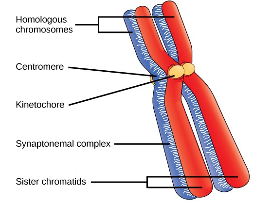
Figure
1.1. Early in prophase I, homologous chromosomes come together to
form a synapse. The chromosomes are bound tightly together and in perfect
alignment by a protein
lattice called a synaptonemal complex
and
by cohesin
proteins at the centromere.
Located at intervals along the synaptonemal complex
are large protein assemblies called recombination
nodules. These assemblies mark the points of later chiasmata and mediate
the multistep process of crossover—or genetic
recombination—between the non-sister chromatids. Near the recombination nodule
on each chromatid, the double-stranded
DNA is cleaved, the cut ends are modified, and a new connection is made between
the non-sister chromatids. As prophase I
progresses, the synaptonemal complex begins to break down and the chromosomes
begin to condense. When the synaptonemal complex is gone, the homologous
chromosomes remain attached to each other at the centromere and at chiasmata.
The chiasmata remain until anaphase I. The number of chiasmata varies according
to the species and the length of the chromosome. There must be at least
one chiasma per chromosome for proper separation of homologous chromosomes during meiosis I, but there may be as many as 25. Following
crossover, the synaptonemal complex
breaks down and the cohesin connection between
homologous pairs is also removed. At the end of prophase I, the pairs are held
together only at the chiasmata (Figure 1.2)
and are called tetrads because the
four sister chromatids of each pair of homologous chromosomes are now visible.
The crossover events
are the first
source of genetic
variation in the nuclei produced
by meiosis. A single crossover event between homologous non-sister chromatids leads to a reciprocal exchange of equivalent DNA between a maternal
chromosome and a paternal chromosome. Now,
when that sister chromatid is moved into a gamete cell it will carry some DNA
from one parent of the individual and some DNA from the other parent. The
sister recombinant chromatid has a combination of maternal and paternal genes
that did not exist before the crossover.
Multiple crossovers in an arm of the chromosome have the same effect, exchanging segments of DNA to
create recombinant chromosomes.
Figure 1.2 Crossover occurs
between non-sister chromatids of homologous chromosomes. The result is an exchange
of genetic material between homologous
chromosomes.
Prometaphase I
The key event in prometaphase I is the attachment of the spindle
fiber microtubules to the kinetochore proteins at the
centromeres. Kinetochore proteins are multiprotein complexes that bind the
centromeres of a chromosome to the microtubules of the mitotic spindle.
Microtubules grow from centrosomes placed at opposite poles of the cell. The
microtubules move toward the middle of the cell and attach to one of the two
fused homologous chromosomes. The microtubules attach at each chromosomes'
kinetochores. With each member of the
homologous pair attached to opposite poles of the cell, in the next phase, the microtubules can pull the homologous pair apart. A spindle fiber that has attached
to a kinetochore is called a kinetochore microtubule. At the end of
prometaphase I, each tetrad is attached to microtubules from both poles, with one homologous chromosome facing each pole. The homologous chromosomes are still held together
at chiasmata. In addition, the nuclear membrane has broken
down entirely.
Metaphase I
During metaphase I, the homologous chromosomes are arranged
in the center of the cell with the kinetochores facing opposite poles. The
homologous pairs orient themselves randomly at the equator. For example, if the two homologous
members of chromosome 1 are labeled a and b, then the chromosomes could line up
a-b, or b-a. This is important in determining
the genes carried
by a gamete, as each will only receive one of the two homologous chromosomes. Recall
that homologous chromosomes are not identical. They contain slight differences in their genetic information,
causing each gamete to have a unique
genetic makeup.
This randomness is the physical
basis for the creation of the second
form of genetic
variation in offspring. Consider that the homologous chromosomes
of a sexually reproducing organism
are originally inherited as two separate sets, one from each parent. Using
humans as an example, one set of 23 chromosomes is present in the egg donated
by the mother. The father provides
the other set of 23 chromosomes in the sperm that fertilizes the egg. Every
cell of the multicellular offspring has copies of the original
two sets of homologous chromosomes. In prophase I of meiosis,
the homologous chromosomes form the tetrads. In metaphase
I, these pairs line up at the midway point between the two poles of the cell
to form the metaphase plate. Because there is an equal chance that a
microtubule fiber will encounter a maternally or paternally inherited chromosome, the arrangement of the tetrads
at the metaphase plate is random. Any maternally inherited chromosome may face either
pole. Any paternally inherited chromosome may also face either pole. The
orientation of each tetrad is
independent of the orientation of the other 22 tetrads.
This event—the random (or independent) assortment of
homologous chromosomes at the metaphase plate—is the second mechanism that introduces variation
into the gametes
or spores. In each cell that undergoes meiosis,
the arrangement of the
tetrads is different. The number of variations is dependent on the number
of chromosomes making
up a set. There are two possibilities for orientation at the metaphase
plate; the possible
number of alignments therefore equals 2n, where n
is the number of chromosomes per set. Humans have 23 chromosome pairs,
which results in over eight million (223)
possible genetically-distinct gametes. This number does not include the
variability that was previously created in the sister chromatids by crossover. Given these two mechanisms, it is highly unlikely that any two haploid cells resulting from meiosis
will have the same genetic
composition (Figure 1.3).
To summarize the genetic consequences of meiosis I, the maternal and
paternal genes are recombined by crossover events that occur between each homologous pair during prophase
I. In addition, the random assortment of tetrads on the metaphase plate produces a unique combination
of maternal and paternal chromosomes that will make their
way into the gametes.
Figure
1.3 Random,
independent assortment during metaphase I can
be demonstrated by
considering a cell with a set of two chromosomes (n =
2). In this case, there
are two possible arrangements at
the equatorial plane in metaphase
I. The total possible number of
different gametes
is 2n, where n equals the number of
chromosomes in a set. In this example, there are four possible genetic combinations for the gametes. With n = 23 in human cells,
there are over 8 million
possible combinations of paternal
and maternal chromosomes.
Anaphase I
In anaphase I, the microtubules pull the linked chromosomes apart. The sister chromatids remain tightly bound together
at the centromere. The chiasmata are broken in anaphase I as the microtubules
attached to the fused kinetochores pull the homologous chromosomes apart (Figure 1.4).
Telophase I and Cytokinesis
In telophase, the separated chromosomes arrive at opposite
poles. The remainder of the typical
telophase events may or may not occur, depending on the species. In some organisms, the chromosomes decondense and nuclear envelopes form
around the chromatids in telophase I. In other organisms, cytokinesis—the physical separation of the cytoplasmic
components into two daughter cells—occurs without reformation of the nuclei. In
nearly all species of animals and some fungi, cytokinesis separates the cell contents
via a cleavage furrow (constriction of the actin
ring that leads
to cytoplasmic division). In plants, a cell
plate is formed during cell cytokinesis by Golgi vesicles fusing at the
metaphase plate. This cell plate will ultimately lead to the formation of cell
walls that separate the two daughter cells.
Two haploid cells are the end result of the first meiotic
division. The cells are haploid
because at each pole, there is just one of each pair of the homologous
chromosomes. Therefore, only one full set of the chromosomes is present. This
is why the cells are considered haploid—there is only one chromosome set, even
though each homolog still consists of two sister chromatids. Recall that sister
chromatids are merely duplicates of one of the two homologous chromosomes
(except for changes that occurred during crossing over). In meiosis II, these
two sister chromatids will separate, creating four haploid daughter cells.
Meiosis II
In some species,
cells enter a brief interphase, or interkinesis, before entering meiosis
II. Interkinesis lacks an S phase,
so chromosomes are not duplicated. The two cells produced in meiosis I go
through the events of meiosis II in synchrony.
During meiosis II, the sister chromatids within the two daughter cells
separate, forming four new haploid gametes. The mechanics of meiosis II is
similar to mitosis, except that each dividing cell has only one set of
homologous chromosomes. Therefore, each
cell has half the number of sister chromatids to separate out as a
diploid cell undergoing mitosis.
Prophase II
If the chromosomes decondensed in telophase
I, they condense again. If nuclear envelopes
were formed, they fragment into vesicles. The centrosomes that were
duplicated during interkinesis move away from each other toward opposite poles,
and new spindles are formed.
Prometaphase II
The nuclear envelopes are completely broken down, and
the spindle is fully formed. Each sister chromatid forms an individual kinetochore that attaches to
microtubules from opposite poles.
Metaphase II
The sister chromatids are maximally condensed and
aligned at the equator of the cell.
Anaphase II
The sister chromatids are pulled apart by the
kinetochore microtubules and move toward opposite poles. Non-kinetochore
microtubules elongate the cell.
Figure
1.4 The process of chromosome alignment
differs between meiosis I and meiosis II. In prometaphase I,
microtubules
attach to the fused
kinetochores of homologous chromosomes, and the homologous chromosomes are
arranged at the midpoint
of the cell in metaphase I.
In anaphase I, the homologous chromosomes are separated. In prometaphase
II, microtubules attach to
the kinetochores of sister chromatids, and the sister chromatids are arranged at the midpoint
of the cells in metaphase II. In anaphase II, the sister
chromatids are separated.
Telophase II and Cytokinesis
The chromosomes arrive at opposite poles and begin to
decondense. Nuclear envelopes form around the chromosomes. Cytokinesis
separates the two cells into four unique haploid cells. At this point, the newly
formed nuclei are both haploid. The cells produced are genetically unique
because of the random assortment of paternal and maternal homologs and because of the recombining of maternal and paternal segments
of chromosomes (with their sets of genes) that occurs during
crossover. The entire process of meiosis is outlined in Figure 1.5.
Figure
1.5 An animal cell with a diploid
number of four (2n = 4) proceeds through the stages of meiosis to
form four haploid daughter cells.
Comparing Meiosis and Mitosis
Mitosis and meiosis are both forms of division of the
nucleus in eukaryotic cells. They share some similarities, but also exhibit distinct
differences that lead to very different outcomes
(Figure 1.6). Mitosis
is a single nuclear division
that results in two nuclei
that are usually partitioned into two new cells. The nuclei resulting from a
mitotic division are genetically identical to the original nucleus. They have
the same number of sets of chromosomes, one set in the case of haploid cells
and two sets in the case of diploid cells. In most plants and all animal
species, it is typically diploid cells that undergo mitosis to form new diploid cells. In contrast, meiosis
consists of two nuclear divisions resulting in four nuclei that are usually
partitioned into four new cells. The nuclei resulting from meiosis are not
genetically identical and they contain one chromosome set only. This is
half the number of chromosome sets
in the original cell, which is diploid.
The main differences
between mitosis and meiosis occur in meiosis I, which is a very different nuclear division than mitosis. In meiosis I, the homologous chromosome pairs become associated with each other, are bound together
with the synaptonemal complex, develop chiasmata and undergo crossover between sister chromatids,
and line up along the metaphase plate in tetrads with kinetochore fibers from opposite
spindle poles attached
to each kinetochore of a homolog in a tetrad. All of these events occur only in meiosis I.
When the chiasmata resolve and the tetrad is broken up
with the homologs moving to one pole or another,
the ploidy level—the number of sets of chromosomes in each future nucleus—has
been reduced from two to one. For this reason, meiosis I is referred to as a reduction division.
There is no such reduction in ploidy
level during mitosis.
Meiosis II is much more analogous to a mitotic
division. In this case, the duplicated chromosomes (only one set of them) line
up on the metaphase plate with divided kinetochores attached to kinetochore
fibers from opposite poles. During anaphase II, as in mitotic
anaphase, the kinetochores divide and one sister chromatid—now referred to as a chromosome—is pulled to one pole while the other sister chromatid is pulled to
the other pole. If it were not for the fact that there had been crossover, the two products of each individual
meiosis II division would be identical (like in mitosis). Instead, they are different because there has always been at
least one crossover per chromosome. Meiosis II is not a reduction division
because although there are fewer copies of the genome in the resulting cells,
there is still one set of chromosomes, as there was at the end of meiosis I.

Figure
1.6 Meiosis and mitosis are both preceded by
one round of DNA
replication; however, meiosis includes
two nuclear divisions. The four daughter cells resulting from meiosis are haploid
and genetically distinct. The daughter cells
resulting from mitosis are diploid and identical to the parent cell.