The
length of the cell cycle is highly variable, even within the cells of a single
organism. In humans, the frequency
of cell turnover ranges from a few hours in early embryonic development, to an average of two to five days for epithelial cells, and to an
entire human lifetime spent in G0 by
specialized cells, such as cortical neurons or cardiac muscle cells. There is
also variation in the time that a cell spends in each phase of the cell cycle. When fast-dividing mammalian cells are grown in culture (outside the body under optimal
growing conditions), the length of the cycle is about 24 hours. In rapidly
dividing human cells with a 24-hour
cell cycle, the G1 phase lasts approximately nine hours, the S phase
lasts 10 hours,
the G2 phase
lasts about four and one-half hours, and the M phase lasts approximately
one-half hour. In early embryos of
fruit flies, the cell cycle is completed in about eight minutes. The timing of
events in the cell cycle is controlled by mechanisms that are both internal and external to the cell.
Regulation of the Cell Cycle
by External
Events
Both
the initiation and inhibition of cell division are triggered by events external
to the cell when it is about to begin the replication process. An event may be
as simple as the death of a nearby cell or as sweeping as the release of
growth- promoting hormones, such as human growth hormone (HGH). A lack of HGH
can inhibit cell division, resulting in dwarfism, whereas too much HGH can
result in gigantism. Crowding of cells can also inhibit cell division. Another
factor that can initiate cell division is the size of the cell; as a cell
grows, it becomes inefficient due to
its decreasing surface-to- volume ratio. The solution to this problem is to divide.
Whatever
the source of the message, the cell receives the signal, and a series of events
within the cell allows it to proceed into interphase. Moving forward from this
initiation point, every parameter required during each cell cycle phase must be
met or the cycle cannot progress.
Regulation at Internal Checkpoints
It
is essential that the daughter cells produced be exact duplicates of the parent
cell. Mistakes in the duplication or distribution of the chromosomes lead to mutations
that may be passed forward
to every new cell produced
from an abnormal cell. To
prevent a compromised cell from continuing to divide, there are internal
control mechanisms that operate at three main
cell cycle checkpoints. A checkpoint is one of several points in the eukaryotic cell cycle at which the progression of a cell to the next stage in the cycle can be halted until conditions are favorable. These checkpoints occur near the end of G1,
at the G2/M transition, and during metaphase (Figure 1).

Figure 1 The cell cycle is controlled at three checkpoints. The integrity
of the DNA is assessed at the G1 checkpoint. Proper chromosome duplication is assessed at the G2 checkpoint. Attachment of each kinetochore to a spindle fiber is assessed at the M checkpoint.
The G1 Checkpoint
The
G1 checkpoint determines whether all
conditions are favorable for cell division to proceed. The G1 checkpoint, also called the
restriction point (in yeast), is a point at which the cell irreversibly commits
to the cell division process. External influences, such as growth factors, play
a large role in carrying the cell
past the G1 checkpoint. In addition to adequate
reserves and cell size, there
is a check for genomic
DNA damage at the G1 checkpoint. A cell that does not meet all the requirements will not be allowed
to progress into the S phase. The cell can halt the cycle and attempt to remedy
the problematic condition, or the cell can advance into G0 and
await further signals when conditions improve.
The G2 Checkpoint
The G2 checkpoint bars entry into the mitotic phase if certain conditions
are not met. As at the G1 checkpoint, cell size
and protein reserves are assessed.
However, the most important
role of the G2 checkpoint is to ensure that all of the chromosomes have been replicated and
that the replicated DNA is not damaged. If the checkpoint mechanisms detect
problems with the DNA, the cell cycle is halted, and the cell attempts to
either complete DNA replication or repair the damaged DNA.
The M Checkpoint
The M checkpoint occurs near the end of the metaphase
stage of karyokinesis. The M checkpoint is also known as
the spindle checkpoint, because it determines whether all the sister chromatids
are correctly attached to the spindle microtubules. Because the separation of
the sister chromatids during anaphase is an irreversible step, the cycle will
not proceed until the kinetochores of each pair of sister chromatids are firmly
anchored to at least two spindle fibers arising from opposite poles of the cell.
Regulator Molecules of the Cell Cycle
In
addition to the internally controlled checkpoints, there are two groups of
intracellular molecules that regulate the cell cycle. These regulatory molecules
either promote progress
of the cell to the next phase (positive regulation) or halt the cycle
(negative regulation). Regulator molecules may act individually, or they can influence the activity or
production of other regulatory proteins. Therefore, the failure of a single
regulator may have almost no effect
on the cell cycle, especially if more than one mechanism controls
the same event.
Conversely, the effect of a deficient or non-functioning regulator can be wide-ranging and possibly fatal to the cell if
multiple processes are affected.
Positive Regulation of the Cell Cycle
Two groups of proteins, called cyclins and cyclin-dependent kinases (Cdks), are responsible for the progress
of the cell through the various checkpoints. The levels of the four cyclin
proteins fluctuate throughout the cell cycle in a predictable pattern (Figure 2).
Increases in the concentration of cyclin proteins are triggered by both
external and internal signals. After the cell
moves to the next stage of
the cell cycle, the cyclins that were active in
the previous stage are degraded.
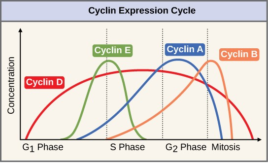
Figure 2 The concentrations of cyclin
proteins change throughout the
cell cycle. There is a
direct
correlation between cyclin accumulation and the three major
cell cycle checkpoints. Also note the sharp decline
of cyclin levels following each checkpoint (the
transition
between phases of the cell
cycle), as cyclin is degraded by cytoplasmic
enzymes. (credit: modification of work by "WikiMiMa"/Wikimedia Commons)
Cyclins
regulate the cell cycle only when they are tightly bound to Cdks. To be fully active, the Cdk/cyclin complex
must also be phosphorylated in specific
locations. Like all kinases, Cdks are enzymes
(kinases) that phosphorylate other proteins.
Phosphorylation activates the protein by changing its shape. The proteins phosphorylated by Cdks are involved in advancing
the cell to the next phase. (Figure 3). The levels of Cdk proteins
are relatively stable throughout the cell cycle; however, the concentrations of cyclin fluctuate and determine when Cdk/cyclin complexes form. The different cyclins
and Cdks bind at specific points in the cell cycle and thus regulate different
checkpoints.

Figure 3 Cyclin-dependent kinases
(Cdks)
are protein kinases that, when fully activated,
can phosphorylate and
thus activate other proteins that advance the cell cycle past
a checkpoint. To become fully activated, a Cdk must
bind to a cyclin protein
and then be phosphorylated by another kinase.
Since
the cyclic fluctuations of cyclin levels are based on the timing of the cell
cycle and not on specific events, regulation of the cell cycle usually occurs
by either the Cdk molecules alone or the Cdk/cyclin complexes. Without a specific concentration of fully activated cyclin/Cdk complexes, the cell
cycle cannot proceed through
the checkpoints.
Although the cyclins are the main regulatory molecules
that determine the forward momentum
of the cell cycle, there are several other mechanisms that
fine-tune the progress of the cycle with negative, rather than positive, effects. These mechanisms essentially block
the progression of the cell cycle until problematic conditions are resolved.
Molecules that prevent the full activation of Cdks are called Cdk inhibitors. Many of these
inhibitor molecules directly
or indirectly monitor a particular cell cycle event.
The block placed on Cdks by inhibitor molecules will not be removed until
the specific event that the inhibitor monitors
is completed.
Negative Regulation of the Cell Cycle
The
second group of cell cycle regulatory molecules are negative regulators.
Negative regulators halt the cell cycle. Remember that in positive regulation, active
molecules cause the cycle to progress. The best understood negative regulatory molecules
are retinoblastoma
protein (Rb), p53, and p21. Retinoblastoma proteins are a group of tumor-suppressor proteins common in many cells.
The 53 and 21 designations refer to the functional
molecular masses of the proteins
(p) in kilodaltons. Much of what is known about cell cycle regulation comes from research conducted with cells that have lost regulatory control.
All three of these regulatory proteins were discovered to be damaged or non-functional in cells that had begun to replicate uncontrollably (became cancerous). In each case, the main cause of the
unchecked progress through the cell cycle
was a faulty copy of the regulatory protein.
Rb, p53, and p21 act primarily
at the G1 checkpoint. p53 is a multi-functional protein
that has a major impact on the commitment of a cell to division
because it acts when there is damaged DNA in cells that are undergoing the preparatory processes during G1. If damaged DNA is detected, p53 halts the
cell cycle and recruits enzymes to repair the DNA. If the DNA cannot be repaired,
p53 can trigger apoptosis, or cell suicide,
to prevent the duplication of damaged chromosomes. As p53 levels rise, the production of p21 is triggered. p21 enforces the halt in the cycle dictated by p53 by binding to and inhibiting the activity of the
Cdk/cyclin complexes. As a cell is exposed to more stress, higher levels of p53
and p21 accumulate, making it less
likely that the cell will move
into the S phase.
Rb
exerts its regulatory influence on other positive regulator proteins. Chiefly, Rb monitors cell size. In the active,
dephosphorylated state, Rb binds to proteins called transcription factors,
most commonly, E2F (Figure 4). Transcription
factors “turn on” specific genes, allowing the production of proteins encoded
by that gene. When Rb is bound to E2F,
production of proteins necessary for the G1/S
transition is blocked.
As the cell increases in size, Rb is slowly
phosphorylated until it becomes inactivated. Rb releases E2F, which can now turn on the gene that
produces the transition protein, and this particular block is removed. For the
cell to move past each of the checkpoints, all positive regulators must be
“turned on,” and all negative regulators must be “turned off.”

Figure 4 Rb halts the cell cycle and releases its hold in response to cell growth.
Rb and other proteins that negatively regulate
the cell cycle are
sometimes called
tumor suppressors.
Why
do you think the name tumor suppressor might be appropriate for these proteins?


No comments:
Post a Comment